“Healing is a matter of time, but it is sometimes also a matter of opportunity”
Welcome back to one of the best dental blogs in the world – Dentalfry.com!
I am Dr.Anooja Chandran from India, doing my final semester of my Master’s in Dental Surgery. Today, I will be giving you a complete explanation of the Treatment of Traumatized tooth.
I am writing these dental notes mainly for students to understand the basics. I am not someone who promotes learning something from a textbook without understanding the basics.
So I tried my best to make all the notes simple and easy to understand. 😀
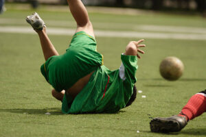
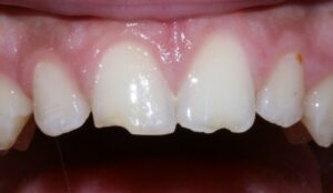
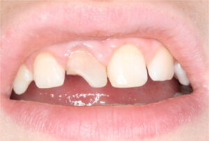
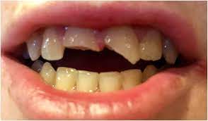
The trauma of the head and neck is commonly seen and constitutes 5% of all dental injuries. It mainly includes the crown fracture and the luxation of the tooth. Consider a situation where you had fallen while playing football.
It can cause a fracture to the crown or root, displacement of the tooth, injury can affect the pulp, crown root fracture, and many more. If the pulp is infected it may recover or survive the damage or in some cases it may undergo degenerative changes or it may finally die or necrotize.
Contents
Cause and Incidence of Dental Injuries
Traumatic injuries to teeth can occur at any age. They are most commonly seen in age groups 2-4 yrs and 8-12 yrs. Boys are more commonly seen than girls.
- Young children falling while playing or walking can be subject to injuries to the front tooth.
- Traumatic injuries in sports and fights are mainly seen in teenagers and adults.
- Child abuse can also cause traumatic injuries to the tooth and the soft tissues.
- Traumatic injuries during bike accidents can be seen in all age groups.
- Domestic violence.
From a dental point of view, they are commonly seen in cases with:
- Protrusion of the anterior teeth.
- Increased overjet.
- Insufficient lip closure.
Fracture of Teeth
| Craze line: Microfracure confined to the enamel. | 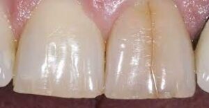 |
| Cuspal fracture: Diagonal fractures that usually do not involve the pulp directly | 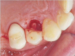 |
| Cracked teeth: Incomplete vertical fracture often involving the pulp | 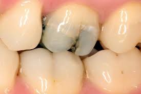 |
| Split tooth: Complete vertical fracture | 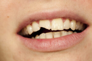 |
| Vertical root fracture: Longitudinal complete fracture usually of endodontically treated teeth. | 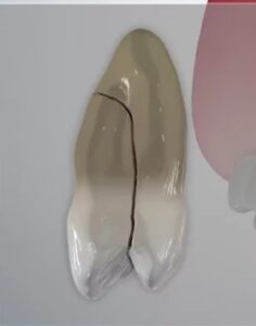 |
Diagnosis of Traumatic Dental Injuries
Clinical Examination
When we diagnose a case of traumatic injury we have to ask the patient about the history, visual examination, Intraoral examination, extra oral examination, electric pulp test, and proper radiograph. The electric pulp test shows negative in the first 3 months because the nerve vessels will be paralyzed or torn out. On clinical examination, you will be able to see slight discoloration of the tooth usually seen in pink color.
It will disappear on its own when the tooth comes to normal. Take multiple angulations of the tooth because the horizontal and diagonal fractures can be determined only by this. CBCT also has to be taken then we will come to know the depth of the fracture.
Radiographic Examination
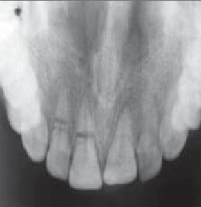
Radiographs have to be taken in more than one angulation. Check for any tooth fragments in the soft tissues, tongue, and buccal mucosa that may get embedded.
- 90-degree horizontal angulation.
- occlusal view.
- lateral view from the mesial and distal aspects.
Enamel Infraction
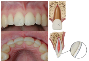
An incomplete fracture of the enamel without fracture of the tooth.
Clinical feature
Clinical examination can be done using dyes or illumination. In transillumination when the light passes through the tooth and sees a fractured line the light will bend and will not pass through the fracture and the opposite tooth looks like dark in color. Tenderness on percussion is commonly negative and if so then check for luxation.
Radiographic feature
No radiographic abnormalities.
Treatment
If the infraction is marked then use etchant and sealing with resin to prevent the infraction. otherwise, no treatment is required.
Enamel Fracture
Enamel fracture is defined as the complete fracture of the enamel.
Clinical feature
On clinical examination, the enamel will be visible. The dentin will not be affected. There will not be any tender on percussion. Check for mobility.
Radiographic examination
On radiographic examination, there will be enamel loss. Radiographs should be taken in three different angulations. Also, check for any fragments in the lip and the cheek.
Treatment
The chipped enamel might make the tooth rough which causes problems in aesthetics. So you can do smoothening of the rough surface and composite restorations can be done.
Enamel Dentin Fracture Without Pulp Exposure
In such cases, there will be a fracture to the enamel and dentin and there won’t be exposure to the pulp.
Clinical feature
As the pulp is not involved there won’t be any tenderness on percussion, if there is so then look for the luxation and root fracture. The pulp vitality test shows that it is usually positive.
Radiographic Examination
On radiographic examination, there will be involvement of the enamel and dentin. Always go for occlusal, periapical, and eccentric radiographs to check for root fractures. Lips and cheeks also must be checked for foreign bodies.
Treatment
In case of a fracture of the tooth without exposure to the pulp, check for the remaining dentin thickness. The remaining dentin thickness of 2mm helps to protect the pulp and shows a good prognosis. There will be an inflammatory response as long as there is a vascular supply. Composite restoration is the treatment of choice and if you have fractured fragments then it can be kept back using the dentin bonding agents and composite resins.
Follow up
A proper follow-up has to be taken and a pulp vitality test in each appointment.
Enamel and Dentin fracture involving the pulp
It’s a fracture involving the enamel and a dentin fracture involving the pulp. The tooth with pulp exposure has more chance of survival than the tooth affected with caries. The fracture’s extent and root development stage should be determined.
Clinical feature
The tooth shows normal mobility. No tender on percussion. If tenderness is present then check for mobility, luxation, and any root fracture. The electric pulp test shows sensitivity.
Radiographic feature
As the enamel, dentin, and pulp is involved there is tooth loss seen in the radiograph.
Treatment
For a tooth with pulp exposure, our main aim is to maintain the vitality of the pulp. Four kinds of treatments can be done.
- Direct pulp capping.
- Pulpotomy.
- Pulpectomy.
- Regenerative Endodontics.
Followup
Clinical and radiographic control at 6-8weeks and 1 year.
Crown-Root Fracture without Pulpal Involvement
A fracture involving the enamel, dentin, and cementum without involving the pulp.
Clinical Feature
It is an oblique fracture line that is a few millimeters incisal to marginal gingiva. It looks like a crown fracture but they also involve the root. There will be a tender on percussion and the coronal fragments are usually mobile.
Radiographic feature
The extension of the fracture should be evaluated. Occlusal, horizontal, and lateral view radiographs have to be taken.
Treatment
Emergency treatment includes stabilization of the fragments at the affected sight.
CBCT scan has to be taken which involves the fracture in the horizontal and diagonal plane.
The treatment modalities include removal or reattachment of the fracture.
Wrapping Up
Did this article help you in any way? Then please do share it with your friends and students in need.
If there is anything you feel I must improve in this, do let me know in the comments. I will try my best to make the notes more student-friendly.
Got any questions?
You can comment your queries in comments below and I will reply to your comments and clear your doubts.
